
Application:
- Complete retinal pigment epithelium (RPE) and outer retinal atrophy (cRORA) is the end point of advanced dry age-related macular degeneration (AMD).
- AMD affects approximately 170 million people globally, with prevalence ranging from 2-6% in people over 60 years old, increasing to 10-20% in those over 75 years. Specifically, cRORA affects about 5 million people worldwide and accounts for approximately 20% of legal blindness cases related to AMD, with an annual incidence rate of 1.5-2% in early AMD patients progressing to cRORA.
- The objective quantification of cRORA is required for clinical diagnosis, follow-up, treatment efficacy evaluation, and clinical research.
- Spectral Domain Optical Coherence Tomography (OCT) has become an essential technology to evaluate the macula, since it provides detailed layer-by-layer visualization of the macula’s microstructure, enabling precise measurement of macular thickness, detection of fluid accumulation, and tracking of disease progression
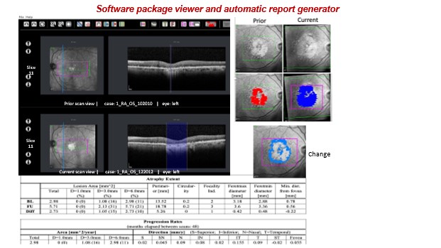
Our innovation:
A new method for the identification and quantification of age-related macular degeneration-end-stage retinal degenerations and dystrophies (cRORA and GA atrophy) in OCT scans and their visualization in the corresponding infrared (IR) image.
The method is based on the classification of light scattering patterns in columns of vertical pixel-wide vectors in OCT slices which atrophy is present with a custom column-based CNN.
The novelties of our method compared to existing ones are:
- accurate classification and segmentation of healthy, incomplete, and complete retinal atrophy in OCT scan slices based on light scattering patterns in column patches
- quantification of the atrophy lesions area, the number of atrophy lesions, and the distance of each atrophy lesion from the fovea,
- visual display of the atrophy lesions computed from the OCT scan onto IR image
Advantages:
- Fully automatic process
- Relies on easy-to-generate OCT atrophy columns for the training set
- Requires significantly fewer manual annotations than existing methods
- User-friendly GUI viewer and editor, “OCT-E”, for the visualization, delineation, and correction of AMD-related retinal atrophy in OCT scans and IR images
- Provides clinically relevant measures for the evaluation of AMD retinal atrophy
Opportunity:
We are seeking collaboration with medical OCT scanner companies providing image analysis software; established EMR, PACS, and HIS companies offering ophthalmology modules; start-up companies providing advanced OCT image processing; or venture capital looking for OCT image analysis technology.